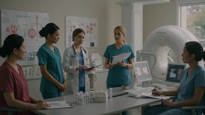
Breast cancer screening and diagnostic techniques are vital for early detection and treatment planning. This comprehensive guide dissects the primary clinical approaches used in the United States to identify, monitor, and diagnose breast cancer, enabling clinicians and patients to make informed decisions. Screening focuses on identifying possible cancer in individuals without symptoms, while diagnostic methods help clarify ambiguous results and determine the exact nature of detected abnormalities.
Understanding these techniques is crucial for American women and healthcare professionals, as regular surveillance can significantly enhance detection rates and potentially impact outcomes. From established imaging tools that lead the field to innovative supplemental technologies, the landscape of breast cancer detection continues to evolve, with improved accuracy and availability.
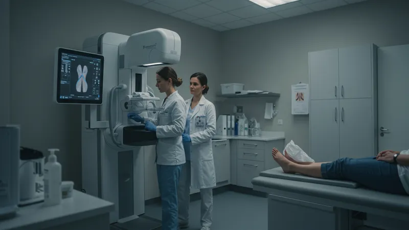
Mammography remains the gold standard for population-wide breast cancer screening in the US, recommended annually or biennially for women within certain age brackets. Its accessibility and relatively low cost have propelled it to the forefront of early detection efforts. However, other screening and diagnostic tools, such as breast MRI and ultrasound, offer valuable alternatives and adjuncts, especially for high-risk individuals or those with dense breast tissue.
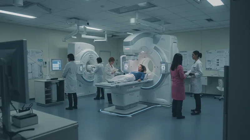
Digital Breast Tomosynthesis, also known as 3D mammography, is transforming the screening landscape by enhancing the visibility of abnormalities in overlapping tissues. Meanwhile, core needle and stereotactic biopsies allow for accurate tissue sampling when imaging results raise suspicion, both playing a pivotal role in confirming diagnoses without requiring surgical procedures. PET scans contribute to staging and monitoring, particularly in complex or advanced cases, although they are not typically used for primary screening due to their cost and specificity.
Alternative imaging modalities—like ductography—address particular diagnostic challenges, such as abnormal nipple discharge, by visualizing the milk ducts directly. Fine needle aspiration provides a less invasive means to sample suspicious lumps, especially useful in outpatient settings to rule in or out malignancy.
Each technique offers unique advantages, limitations, and appropriate contexts for use. Insurance coverage and regional availability can affect access and affordability in the United States, emphasizing the importance of personalized screening strategies that factor in individual risk profiles, breast density, family history, and physician recommendations.
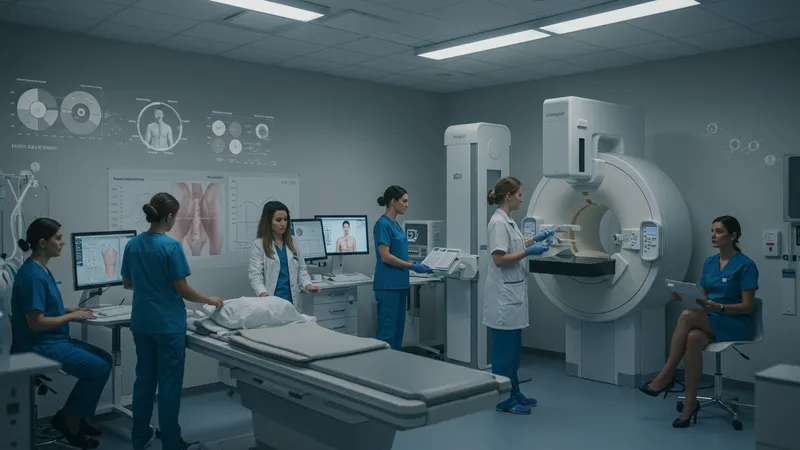
This comprehensive toolkit underscores the dynamic nature of breast cancer detection and diagnosis in the US. The methods detailed above interconnect, forming an optimized pathway from prevention and early identification to confirmation and staging. The deeper details reveal even more valuable insights ahead, including comparative advantages, innovations, and evolving guidelines that drive effective breast health management.
Mammography is the cornerstone of national screening guidelines, with decades of long-term data supporting its effectiveness in reducing breast cancer-related mortality. Designed to detect lesions before they become clinically palpable, this method is accessible at thousands of facilities across the US, often covered in part or in full by health insurance providers under preventive care regulations.
Breast MRI, while more expensive, plays an instrumental role for individuals at substantially elevated risk—such as those with BRCA mutations or strong family history. Its high sensitivity outperforms mammography and ultrasound for detecting certain aggressive tumors, though its lower specificity means it tends to generate more false positive results, sometimes leading to unnecessary biopsies.
Ultrasound is especially valuable as a supplemental tool for characterizing lesions detected on mammography or felt during physical examination. It is non-ionizing, making it suitable for younger women or those requiring repeated imaging. While not a substitute for mammography as a general population screening tool, it increases detection rates in women with dense breasts when used adjunctively.
Digital Breast Tomosynthesis is quickly gaining ground, as emerging research demonstrates its superiority over traditional 2D mammography in detecting invasive cancers and reducing callback rates. However, adoption is variable depending on access, with urban areas more likely to offer the service, sometimes incurring small out-of-pocket expenses that may not be universally covered by insurance plans.
Diagnostic imaging, such as targeted mammography and stereotactic biopsy, is generally reserved for patients who have had abnormal screening results or present with specific symptoms. These advanced imaging strategies enable precise targeting of suspicious areas and enable radiologists to distinguish between benign and malignant changes.
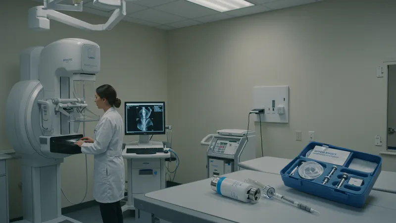
Core needle biopsy remains a minimally invasive method to obtain tissue samples and is widely regarded as the diagnostic standard once imaging alone cannot definitively classify a lesion. The cost for this procedure can vary, but it is routinely performed in outpatient settings and is typically less costly than open surgical biopsies.
Stereotactic biopsy serves a critical role in sampling microcalcifications or small lesions that are difficult to localize on ultrasound. Using real-time X-ray guidance, this technique can be performed swiftly, often negating the need for more invasive surgery. While effective, the expense and access may fluctuate based on geographic region and insurance coverage.
When assessing distant disease spread or monitoring complex cases, PET scans offer unparalleled insights, although they are more commonly used in staging and surveillance. Their high cost limits frequent utilization; however, they are invaluable where detailed whole-body imaging is necessary to guide therapeutic decisions.
The ongoing evolution of breast imaging in the United States is introducing improvements that enhance both detection rates and patient experience. Digital Breast Tomosynthesis has led to more accurate cancer detection, with studies indicating a reduction in false positives and unnecessary biopsies. Availability is increasing, with many centers now integrating 3D mammography into routine screening programs.
Magnetic resonance imaging is seeing technological refinements, including the development of abbreviated MRI protocols. These shorter scans lower costs and increase convenience, making MRI more accessible to broader risk groups. However, insurance coverage may still be limited to those with documented higher risk.
Automated breast ultrasound systems and advances in digital pathology are poised to make screening and diagnosis less dependent on operator skill and more uniform nationwide. These improvements may particularly benefit women with dense breast tissue, which can obscure results on standard mammograms.
Artificial intelligence is beginning to play a supporting role in both screening and diagnosis, offering enhanced algorithms for image interpretation. While regulatory approval and clinical adoption are ongoing processes, these digital tools show potential for further reducing errors and standardizing care across diverse healthcare settings.
Personalizing breast cancer screening in the US involves aligning techniques to each individual’s unique risk profile. Factors such as age, family history, genetic markers, and breast density guide the selection of tools like MRI, tomosynthesis, or supplemental ultrasound. This tailored approach aims to maximize effectiveness while minimizing unnecessary interventions, striving for a higher standard of preventative care.
For individuals with dense breasts—a common challenge, especially in women under 50—combining mammography with ultrasound or tomosynthesis is increasingly recommended. Laws in many US states require providers to inform patients about breast density and its implications for cancer detection, empowering women to make more informed decisions about their care pathways.
Insurance coverage is another essential variable influencing access to advanced diagnostics. While screening mammography is generally covered, additional imaging or biopsies may incur costs depending on plan specifics and patient risk classification. Public and private efforts continue to focus on expanding equitable access and removing financial barriers for high-value clinical testing.
The future of breast cancer screening in the United States is likely to be shaped by emerging technologies, patient-centered care models, and ongoing educational campaigns. Effective integration of these components ensures more comprehensive detection and diagnostic accuracy for American women, equipping patients and clinicians to navigate the evolving landscape of breast health with confidence and clarity.